CT Scan
How should I prepare?
You should wear comfortable, loose-fitting clothing to your exam. You may be given a gown to wear during the procedure.
Metal objects including jewellery, eyeglasses, dentures and hairpins may affect the CT images and should be left at home or removed prior to your exam. You may also be asked to remove hearing aids and removable dental work.
The radiologist also should know if you have diabetes-particularly if you are taking Glucophage.
Women should always inform their physician and the CT technologist if there is any possibility that they are pregnant.
What does the equipment look like?
The CT scanner is typically a large, box like machine with a hole, or short tunnel, in the centre. You will lie on a narrow examination table that slides into and out of this tunnel. Rotating around you, the x-ray tube and electronic x-ray detectors are located opposite each other in a ring, called a gantry. The computer workstation that processes the imaging information is located in a separate room, where the technologist operates the scanner and monitors your examination.
How does the procedure work?
In many ways CT scanning works very much like other x-ray examinations. X-rays are a form of radiation-like light or radio waves-that can be directed at the body. Different body parts absorb the x-rays in varying degrees.
With CT scanning, numerous x-ray beams and a set of electronic x-ray detectors rotate around you, measuring the amount of radiation being absorbed throughout your body. At the same time, the examination table is moving through the scanner, so that the x-ray beam follows a spiral path. A special computer program processes this large volume of data to create two-dimensional cross-sectional images of your body, which are then displayed on a monitor. This technique is called helical or spiral CT.
CT imaging is sometimes compared to looking into a loaf of bread by cutting the loaf into thin slices. When the image slices are reassembled by computer software, the result is a very detailed multidimensional view of the body's interior.
Refinements in detector technology allow new CT scanners to obtain multiple slices in a single rotation. These scanners, called "multislice CT" or "multidetector CT," allow thinner slices to be obtained in a shorter period of time, resulting in more detail and additional view capabilities.
How is the procedure performed?
The technologist begins by positioning you on the CT examination table, usually lying flat on your back or possibly on your side or on your stomach. Straps and pillows may be used to help you maintain the correct position and to hold still during the exam.
Next, the table will move quickly through the scanner to determine the correct starting position for the scans. Then, the table will move slowly through the machine as the actual CT scanning is performed.
When the examination is completed, you will be asked to wait until the technologist verifies that the images are of high enough quality for accurate interpretation.
A CT scan of the head is usually completed within 10 minutes.
What will I experience during and after the procedure?
CT exams are generally painless, fast and easy. With helical CT, the amount of time that the patient needs to lie still is reduced.
If an intravenous contrast material is used, you will feel a slight pin prick when the needle is inserted into your vein. You may have a warm, flushed sensation during the injection of the contrast materials and a metallic taste in your mouth that lasts for a few minutes. Some patients may experience a sensation like they have to urinate but this subsides quickly.
When you enter the CT scanner, special lights may be used to ensure that you are properly positioned. With modern CT scanners, you will hear only slight buzzing, clicking and whirring sounds as the CT scanner revolves around you during the imaging process.
You will be alone in the exam room during the CT scan. However, the technologist will be able to see, hear and speak with you at all times.
After a CT exam, you can return to your normal activities. If you received contrast material, you may be given special instructions.
Who interprets the results and how do I get them?
A physician, usually a radiologist with expertise in supervising and interpreting radiology examinations, will analyse the images and send a signed report to your primary care physician or the physician who referred you for the exam, who will discuss the results with you.
What is CT?
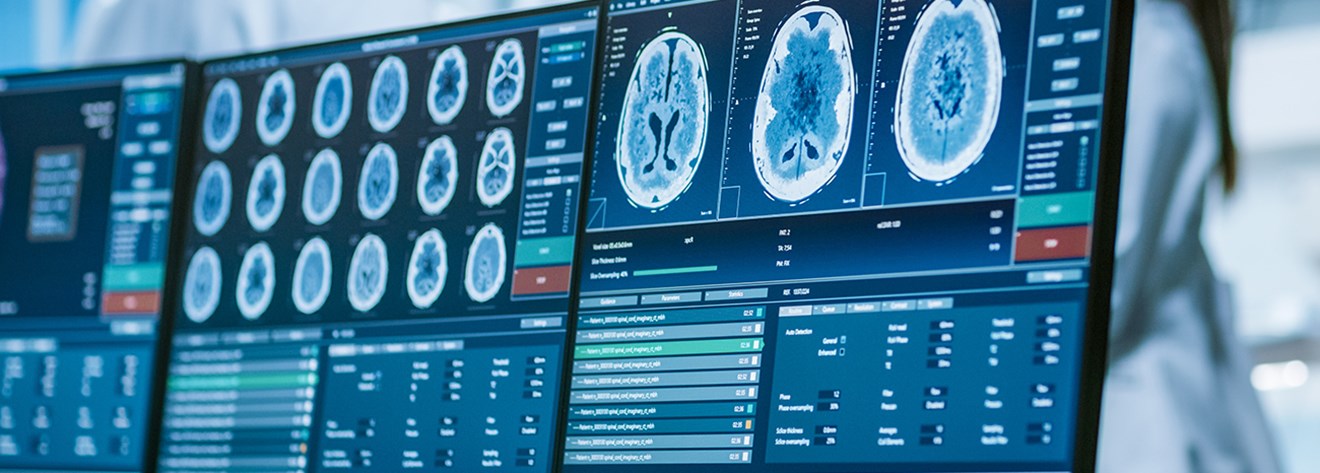
CT scan (or CAT scan) stands for Computerised (Axial) Tomography scan. A CT scanning machine is large and shaped rather like a doughnut and has a couch that you lie on. CT scanning combines special x-ray equipment with sophisticated computers to produce multiple images or pictures of the inside of the body. These cross-sectional images, or "slices" of the area being studied can then be examined on a computer monitor, printed or transferred to a CD.
The Procedure
Unless specifically instructed you may eat and drink as normal, before and after your scan.
If you are just having a CT of your head, you may not be asked to undress. For scanning other parts of the body you may be asked to change into a gown.
You must take off any jewellery in the area to be scanned because metal interferes with the machine.
You will be asked to lie on the scanner table with your head toward the "donut hole" of the CT scanner. The radiographer will position you on the table, and a device to hold your head in place may be used. The radiographer will then leave the room and go to the control room, where you can still communicate by intercom This is because there will be X-rays in the room and it would be dangerous for the staff to be exposed to these all day every day.
An intravenous dye (contrast dye) may be given, through injection. This can help to highlight certain areas of the scan.
While CT images are being taken, it is important to lie still on the table, which will be making a whirring noise during the exam and moving very slowly in order to image the brain or other body part.
Most scans take about half an hour. A lot of that time is to set up the scan, rather than actually taking it
At the end of the scan you may leave and are free to return home.
What happens next?
It can take time for test results to come through. How long will depend on why you are having the scan. Usually, the scan is examined by a specialist in radiology and a report is typed up. The report is then sent to Dr Rose, who will give you the results either when you come to the clinic or by letter which ever was agreed. Please check your clinic letter which will say how you are getting your results.
Does the scan hurt?
The test is painless and there are few side effects. It involves being exposed to some radiation and this is why doctors are careful not to any such scans without good reason. But it isn't a large enough amount of radiation for you to have any ill effects.
We sometimes give a small injection in to a vein in your arm to enhance the scans and give more information. If you receive contrast dye, there is a small chance of an allergic reaction. The reaction most often starts with weakness, sweating and difficulty breathing. The doctors and radiographers will know what to do if you have this type of reaction and will treat you very quickly. If you have allergies to any foods or medicines, particularly seafood or iodine, it is important to inform the technologist before the procedure.
You should also tell the technologist if you could be pregnant as x-ray radiation to the baby is best avoided unless essential.